FLUORESCENCE MICROSCOPY
Super resolution microscope: single molecule localization (SML) based on the microscopy technique SPDM (spectral precision distance microscopy) with up to four different excitation lasers
nanoFLeyeTM
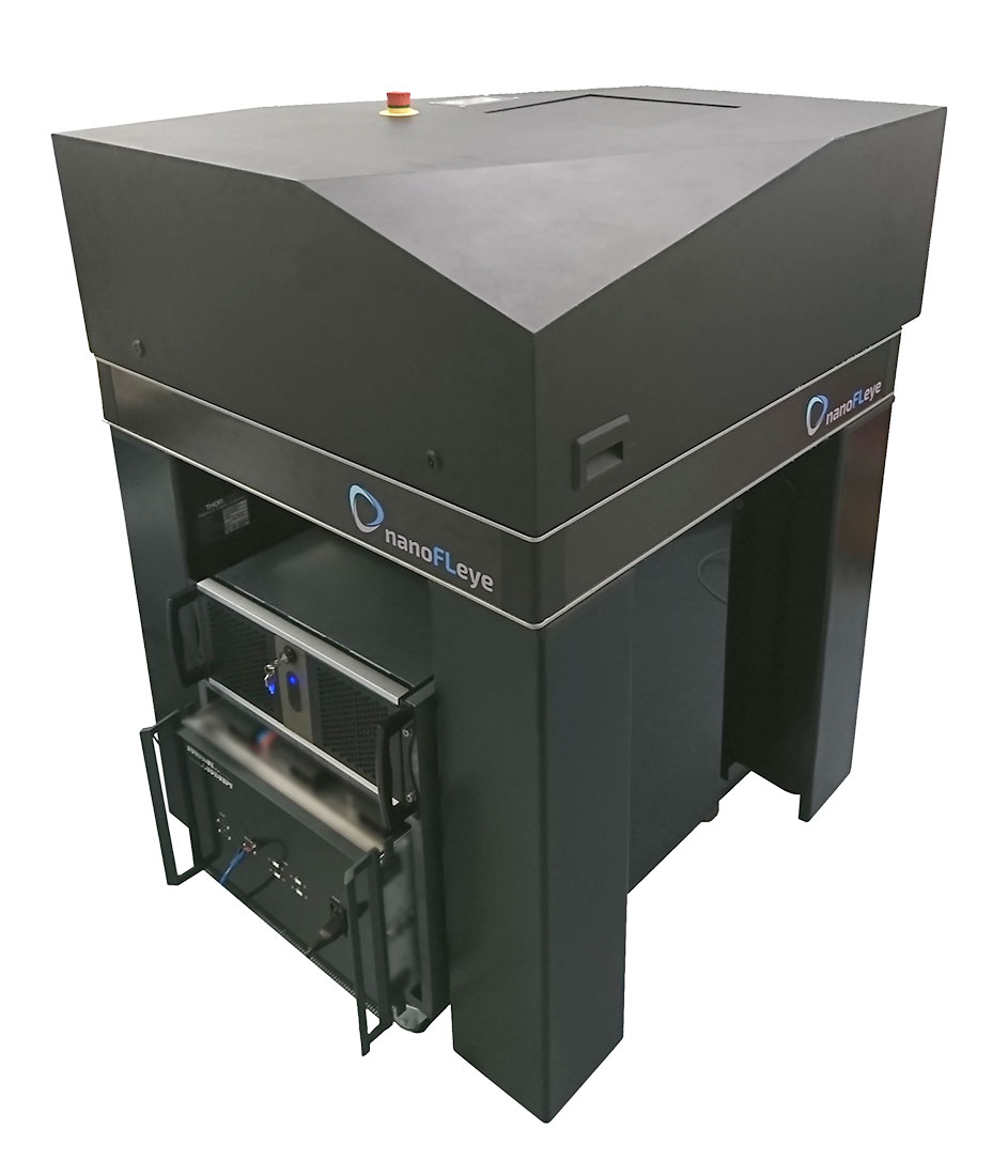
The nanoFLeyeTM is a robust, high-resolution fluorescence microscope that was specially developed for examining the smallest of samples.
- Standard widefield fluorescence microscopy, TIRF microscopy and super resolution based on SPDM
- Movable „All-in-One“ device with dimensions: (width x depth x height) 100x100x130cm
- Multicolour imaging with up to four different laser colours, standard wavelengths available: 405nm, 488nm, 532nm, 561nm and 647nm
For additional information visit nanofleye.com
MORE INFORMATION
In order to guarantee the highest possible flexibility, the nanoFLeyeTM is mounted on a high-quality, cushioned table with integrated PC that can be repositioned and transported without readjustment. It consists of a vertical setup where the specimen is loaded from top. The use of special oil immersions objectives (60x/NA 1.49) enables TIRF microscopy (Total Internal Reflection Fluorescence) in addition to normal widefield imaging and the built-in x,y sample stage for standard specimen slides (76x26mm) and 6-96 well plates (127.7×85.4mm) enables the use of life-cell incubators.
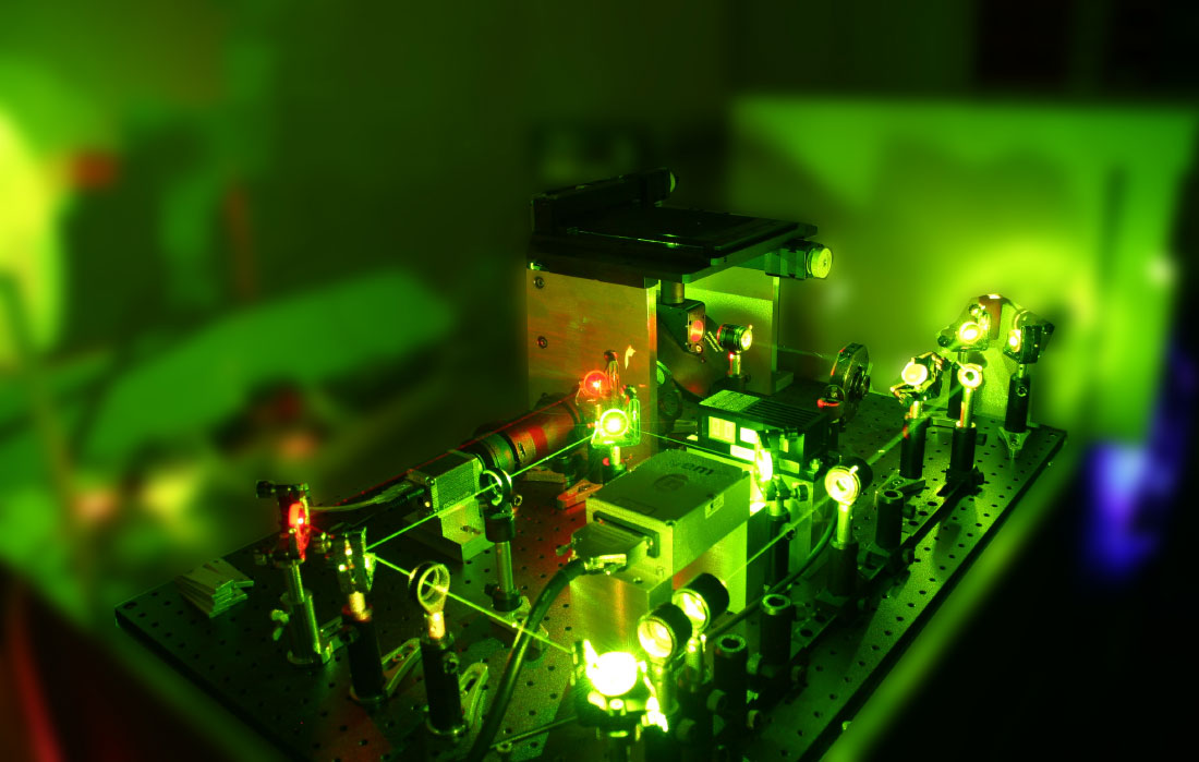
In combination with the specially developed ReconFlexTM camera, excellent images can be created in the shortest possible time. This is made possible by the high speed of the camera. The post-processing time, in case of localization microscopy, is greatly reduced thanks to special integrated algorithms. The high degree of automation simplifies handling and thanks to the modular structure of the device, experienced microscope users can optimize the system for their own experiments and research.
Spectral Precision Distance Microscopy (SPDM): Spectral features are used to achieve optical isolation
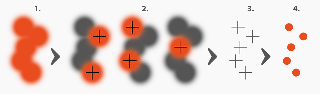
- In Conventional Fluorescence Microscopy the Full-Width-at-Half-Maximum (FWHM) of the Point-Spread-Function (PSF) >200nm. Signals of adjacent dyes overlap, therefore single molecules cannot be resolved
- Using SPDM, randomly activated dyes are „optically isolated“, i.e. no overlap of the signal of adjacent molecules can occur
- The locations of the optically isolated fluorophores are determined by the localization algorithm with a precision down to 20nm
- All localizations found in a stack of usually ten thousands of images are displayed in a single reconstructed super-
resolved image
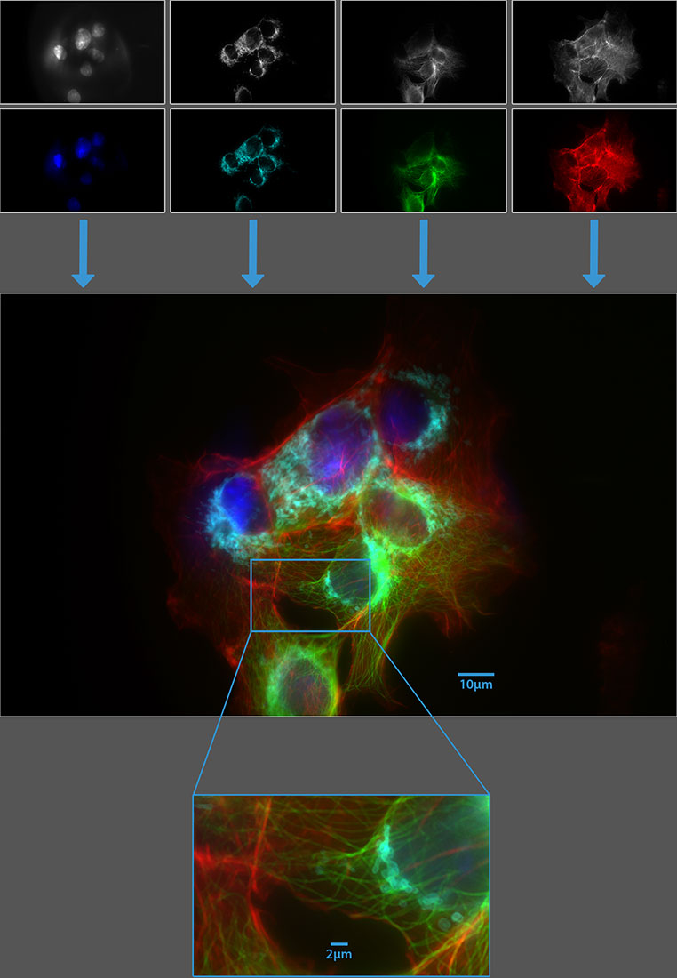
The images show Cos-7-cells recorded via standard widefield fluorescence microscopy.
The different monochrome images (wavelength from left to right: 405nm, 488nm, 561nm, 647nm) where consecutively recorded with a ReconFlexTM 1900.
The cells where stained with DAPI for nucleus (blue), Alexa Fluor 488 for mitochondria (cyan), TMR for microtubules (green) and SiR for actin (red).
The lowest picture shows the four merged channels with an amplified crop.

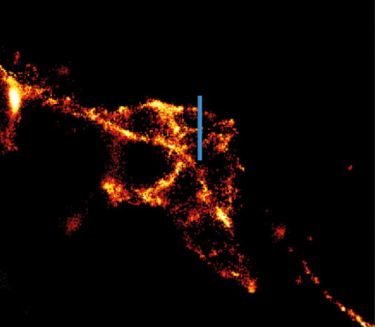
The images show a segment of fluorescence-labeled microtubules in a cell culture (HeLa-cells).
On the left side is a conventional epifluorescent image with a scale bar of 1µm for comparison.
On the right side is a super-resolved image of the identical sample position, in which the algorithm for the super resolution contained in the ReconFlexTM1900 (S)-Camera reveals the substructures.