Time-of-Flight Momentum Microscope With Spin Imaging Option
Momentum microscopy and spectroscopy system: Time-of-Flight and parallel imaging spin analyzers based on the parallel working, patented, Ir or Ir/Au spin filter principle.
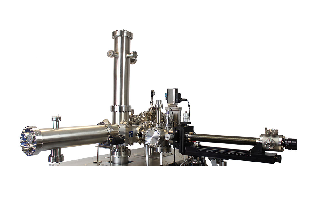
Time-of-Flight Momentum Microscope
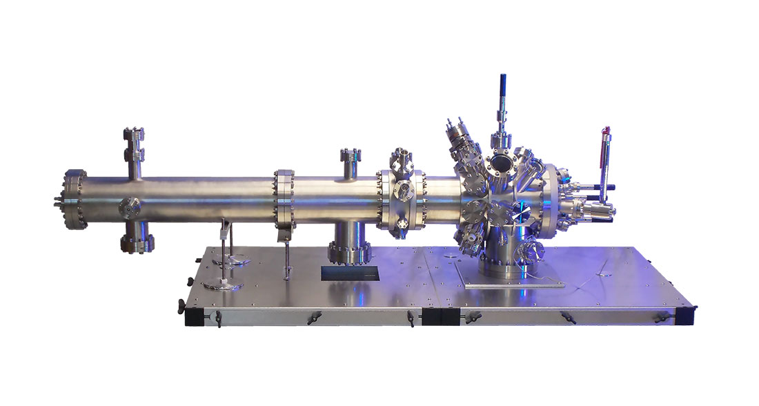
The patented [patents DE102013005173C5 and DE102014018555B3] Time-Of-Flight Momentum Microscope images the full emission hemisphere (2πk²) k-space out of a selectable real space sample area down to a diameter of <1µm, a novel type of ARPES.
- Momentum resolution <0,01Å-1
- Spatial resolution <50nm
- Energy resolution <20meV
- L-He cooled sample stage available
- Parallel spin imaging option available
MORE INFORMATION

The ToF Momentum Microscope works this way: The zoom optics 1 select a real-space sample area, the switching between real-space and k-space-image is done by the zoom optics 2.
[Medjanik et al., Nature Materials, 16(6):615-621(2017)]
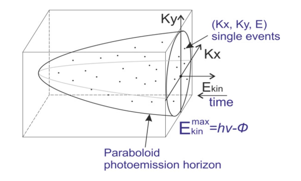
The user can access the full space of the photoemission paraboloid from work function cut-off to Fermi edge during one measurement (approved for excitation energies up to 21.7eV). The optics is isogenic in k for a wide range of energies.
Technical specifications for a complete Momentum Microscope system including the hexapod sample stage.
| ToF Momentum Microscope | |
|---|---|
| Energy resolution | <30meV (guaranteed), <20meV (typical value) |
| Simultaneously focused energy range | Up to 10eV |
| Momentum resolution | <0.01Å-1 (guaranteed) |
| Momentum resolved range | +- 3Å-1 |
| Lateral resolution | <50nm |
| Real space field of view | 11...1000μm |
| Piezo driven contrast aperture | 7 aperture sizes and a 200 mesh, x/y adjustable |
| Piezo driven field aperture | 14 aperture sizes (down to 10μm possible) and a 200 mesh, x/y adjustable |
| Motorized manipulator | 6 axis (Hexapod) which makes in situ sample tilt adjustment possible (e.g. for cleaved samples) |
| Temperature range | <15K....400K (guaranteed), <10K (typical value) |
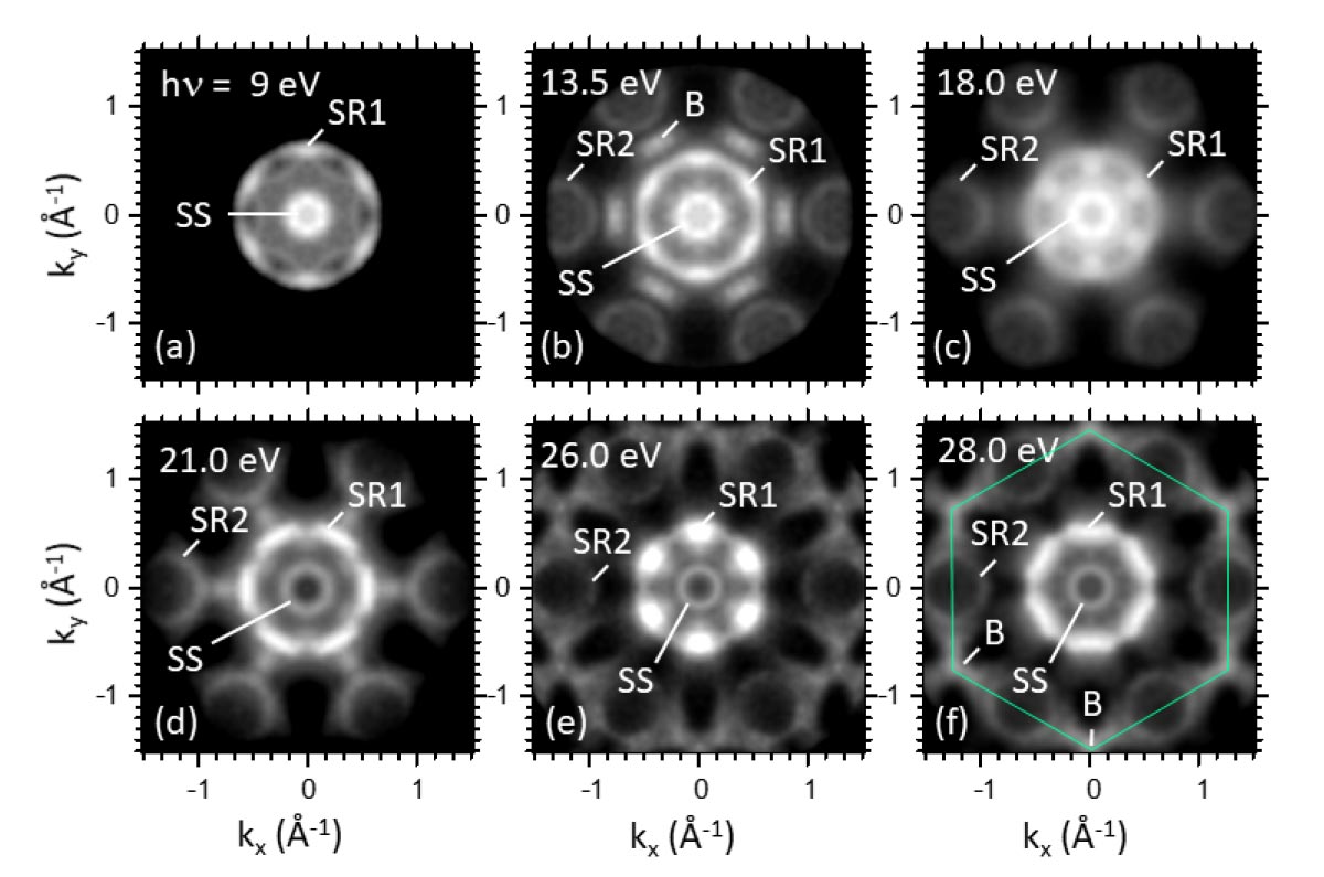
Constant-energy maps measured at various photon energies of Re (0001).
[H.J. Elmers et al. PhysRevResearch.2.013296]
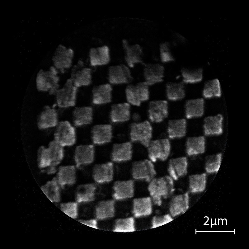
Real space image of an Au on Si (Chessy) sample.
FoV 11µm.
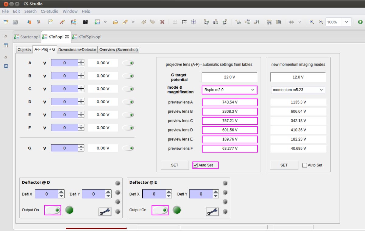
The EPICS (https://epics-controls. org) based software supports fully remote controlled measurements via PC. The server-client architecture enables customized, automatic measurement routines via user scripts.
You can integrate additional customer specific devices by implementing further EPICS modules.
Hexapods for Time-of-Flight Momentum Microscope
The Hexapod is a L-He cooled 6-axis sample stage, reaching temperatures < 15K (lowest value shown < 9K). It enables very precise sample positioning, including rotation and tilt up to ±5°. Each axis is equipped with a position readout. Therefore the user can store sample positions which can be recalled later on.

Imaging Spin Detector for Time-of-Flight Momentum Microscope
The Imaging Spin Filter is an upgrade for an existing ToF Momentum Microscope. The spin imaging is based on a parallel working, patented [patents DE102013005173C5 and DE102005045622B4], Au/Ir spin filter principle.
MORE INFORMATION
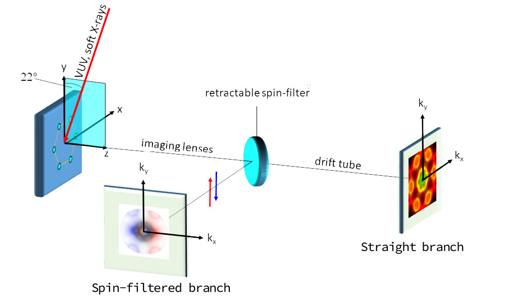
Working principle of the parallel spin analyzer. The instrument works either as a conventional ToF Momentum Microscope in the straight branch or the spin filter crystal deflects the image for spin analysis in the perpendicular branch (spin-filtered branch).
[H.J. Elmers et al., Phys. Rev. Research 2, 013296 (2020)]
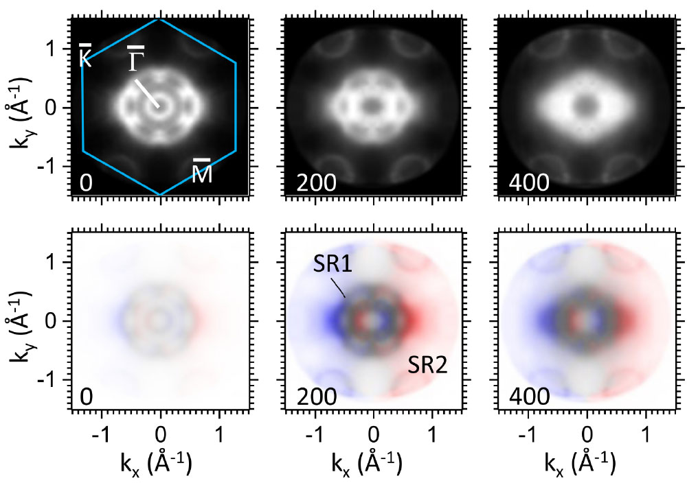
Constant-energy maps of Re (0001) measured with photon energies of 18.5eV. Left to right binding energies 0mV, 200meV and 400meV. The lower row displays the spin filtered measurements.
[H.J. Elmers et al., Phys. Rev. Research 2, 013296 (2020)]

An image with a FoV 450µm of a chessy sample with >9.000 pixels in the complete image. This demonstrates the image quality of the spin-filtered branch.